Nerve Trauma, Entrapment and RSD
Unique characteristics to these conditions
Nerve Trauma, Entrapment, and RSD
Unique Characteristics of These Conditions
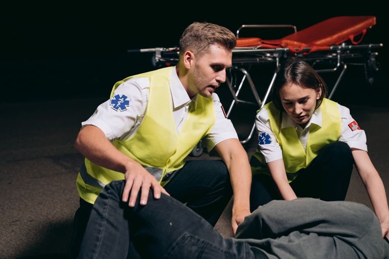
Nerve trauma, nerve entrapment, and Reflex Sympathetic Dystrophy (RSD), also known as Complex Regional Pain Syndrome (CRPS), are serious neurological conditions that can cause chronic pain, weakness, sensory disturbances, and functional limitations. These conditions often develop after injury, surgery, repetitive stress, or trauma affecting the nervous system.
Because nerves control sensation, movement, and communication throughout the body, damage or irritation can lead to symptoms that significantly impact daily life and overall well-being.
Nerve Trauma
Nerve trauma occurs when a nerve becomes injured due to physical damage or excessive stretching, compression, or inflammation.
Common Causes Include:
- Auto accidents
- Slip and fall injuries
- Sports injuries
- Workplace accidents
- Surgical complications
- Lacerations or blunt force trauma
Symptoms of Nerve Trauma May Include:
- Sharp or burning pain
- Numbness or tingling
- Muscle weakness
- Loss of sensation
- Reduced coordination
The severity of symptoms depends on the extent and location of the nerve injury.
Nerve Entrapment
Nerve entrapment occurs when surrounding tissues place pressure on a nerve, disrupting normal nerve function.
Common Entrapment Conditions Include:
- Carpal Tunnel Syndrome
- Cubital Tunnel Syndrome
- Thoracic Outlet Syndrome
- Piriformis Syndrome
- Pudendal Nerve Entrapment
- Radiculopathy
Common Symptoms Include:
- Radiating pain
- Tingling sensations
- Weakness
- Burning discomfort
- Sensitivity to touch

Reflex Sympathetic Dystrophy (RSD) / Complex Regional Pain Syndrome (CRPS)
RSD/CRPS is a chronic pain disorder involving abnormal nerve signaling and dysfunction of the sympathetic nervous system. The condition often develops after an injury, surgery, or trauma and may affect an arm, leg, hand, or foot.
Common Symptoms Include:
- Severe burning pain
- Extreme sensitivity to touch
- Swelling
- Skin color or temperature changes
- Joint stiffness
- Muscle spasms
Pain is often significantly more severe than expected for the original injury.

Unique Characteristics of These Conditions
Chronic Pain
Pain may persist long after the original injury has healed and can become debilitating without proper treatment.
Sensory Disturbances
Patients may experience: Tingling, Numbness, Burning sensations, Hypersensitivity, or Electric shock-like pain.
Functional Limitations
Nerve dysfunction can interfere with: Walking, Lifting, Sitting, Gripping objects, and Daily activities.
Progressive Symptoms
Without treatment, symptoms may worsen over time and lead to long-term nerve damage or disability.
Diagnosis and Evaluation
A comprehensive neurological evaluation is important for identifying the source and severity of nerve dysfunction. Testing includes:
- Physical and neurological examinations
- MRI or CT imaging
- EMG and nerve conduction studies
- Diagnostic nerve blocks
- Functional movement evaluations
Treatment Options
Treatment focuses on relieving pain and improving function. Common Approaches Include:
- Medication management
- Physical & Occupational therapy
- Nerve blocks
- Neuromodulation therapies
- Rehabilitation programs
- Surgical consultation when necessary
Importance of Early Intervention
Early diagnosis and treatment may help prevent worsening nerve damage, improve mobility and strength, reduce chronic pain, and improve overall quality of life. Prompt intervention is especially important for progressive nerve conditions such as RSD/CRPS.
Comprehensive Neurological and Pain Care
Managing nerve trauma, entrapment disorders, and RSD/CRPS requires a personalized and comprehensive approach focused on identifying the underlying cause of symptoms, reducing pain, and improving overall function. Coordinated neurological and rehabilitative care can help patients regain independence and improve long-term outcomes.